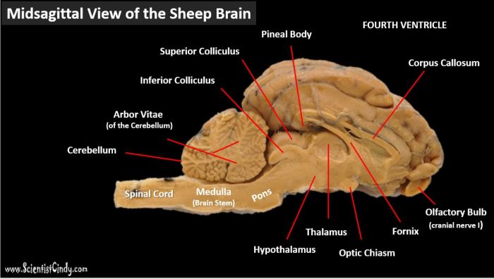
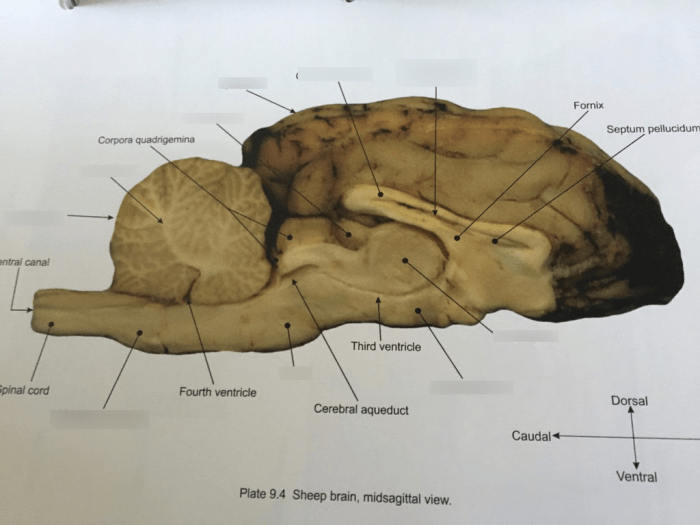
Delve into the intricate depths of the sheep brain midsagittal view labeled, a captivating journey that unveils the remarkable structures and functions of this vital organ. This detailed exploration illuminates the cerebrum, cerebellum, brainstem, ventricular system, meninges, and blood supply, providing an unparalleled understanding of their roles in maintaining life and cognition.
The midsagittal view, a precise section along the brain’s midline, offers a comprehensive perspective of its internal architecture. By examining this view, neuroscientists gain invaluable insights into the brain’s intricate organization and the complex interactions between its various components.
Sheep Brain Midsagittal View Labeled: An Overview

The midsagittal view of the sheep brain is a valuable tool for studying the anatomy and function of the brain. This view provides a clear cross-sectional image of the brain, allowing for the identification of major structures and the examination of their relationships to each other.
The anatomical structures visible in the midsagittal view include the cerebrum, cerebellum, brainstem, and ventricular system. The cerebrum is the largest part of the brain and is responsible for higher-order functions such as cognition, memory, and language. The cerebellum is located behind the cerebrum and is responsible for coordination and balance.
The brainstem connects the cerebrum and cerebellum to the spinal cord and contains vital structures such as the midbrain, pons, and medulla oblongata.
Cerebrum and Cerebellum

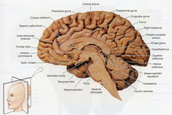
The cerebrum is divided into two hemispheres by the falx cerebri. Each hemisphere is further divided into lobes by sulci (grooves) and gyri (ridges). The major sulci of the cerebrum include the central sulcus, lateral sulcus, and parieto-occipital sulcus. The major gyri of the cerebrum include the precentral gyrus, postcentral gyrus, superior temporal gyrus, and inferior temporal gyrus.
The cerebrum is responsible for a wide range of functions, including cognition, memory, language, and motor control. The cerebellum is responsible for coordination and balance.
Brainstem and Ventricular System: Sheep Brain Midsagittal View Labeled
The brainstem is located inferior to the cerebrum and cerebellum and connects these structures to the spinal cord. The brainstem consists of the midbrain, pons, and medulla oblongata. The midbrain contains structures involved in motor control and eye movements. The pons contains structures involved in sleep and arousal.
The medulla oblongata contains structures involved in vital functions such as breathing and heart rate.
The ventricular system is a series of interconnected cavities within the brain that are filled with cerebrospinal fluid. The ventricular system consists of the lateral ventricles, third ventricle, cerebral aqueduct, fourth ventricle, and central canal.
Meninges and Blood Supply

The meninges are three layers of connective tissue that surround the brain and spinal cord. The meninges include the dura mater, arachnoid mater, and pia mater. The dura mater is the outermost layer and is attached to the skull. The arachnoid mater is the middle layer and is filled with cerebrospinal fluid.
The pia mater is the innermost layer and is closely attached to the surface of the brain.
The brain is supplied with blood by the carotid arteries and the vertebral arteries. The carotid arteries supply blood to the anterior part of the brain, while the vertebral arteries supply blood to the posterior part of the brain.
Clinical Significance
The midsagittal view of the sheep brain is used in diagnosing and treating a variety of brain disorders. For example, this view can be used to visualize tumors, strokes, and other abnormalities in the brain.
The midsagittal view of the sheep brain is a valuable tool for studying the anatomy and function of the brain. This view provides a clear cross-sectional image of the brain, allowing for the identification of major structures and the examination of their relationships to each other.
Essential Questionnaire
What is the significance of the sheep brain midsagittal view?
The sheep brain midsagittal view provides a comprehensive cross-sectional view of the brain, allowing researchers and clinicians to study its internal structures and their relationships in detail.
How is the sheep brain midsagittal view used in clinical practice?
The sheep brain midsagittal view is a valuable tool for diagnosing and treating brain disorders. By comparing a patient’s brain scan to the normal midsagittal view, clinicians can identify abnormalities and determine the underlying cause of neurological symptoms.
What are the major anatomical structures visible in the sheep brain midsagittal view?
The major anatomical structures visible in the sheep brain midsagittal view include the cerebrum, cerebellum, brainstem, ventricular system, meninges, and blood vessels.