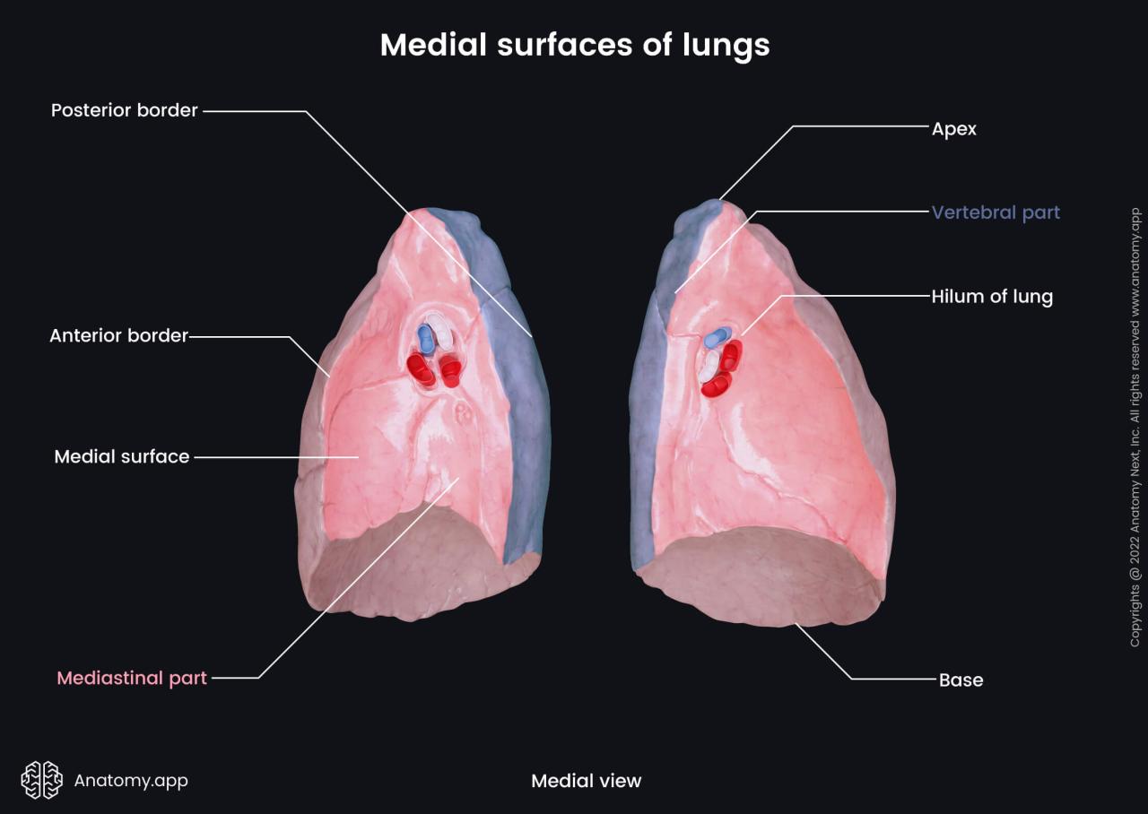
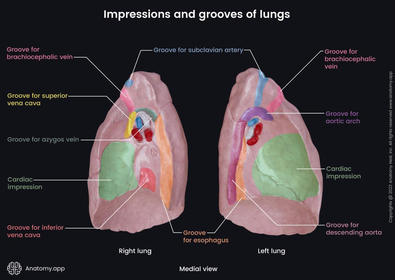
Cardiac impression vs cardiac notch – Delving into the realm of cardiac anatomy, we embark on a journey to decipher the subtle nuances between cardiac impression and cardiac notch. These distinct features, often encountered in medical imaging, hold significant implications for understanding cardiac health.
As we unravel the causes, pathophysiology, and clinical significance of these anatomical landmarks, we’ll equip you with a comprehensive understanding of their diagnostic and therapeutic implications.
Definitions
Cardiac impression and cardiac notch are two distinct anatomical features found on the posterior aspect of the sternum, the breastbone. Both are related to the heart’s position and movement within the chest cavity.
The cardiac impression is a shallow depression on the posterior surface of the sternum, where the heart rests against it. It is usually located at the level of the fourth and fifth costal cartilages, which connect the ribs to the sternum.
The cardiac impression is more prominent in individuals with a thin chest wall and is less noticeable in those with a thick chest wall.
The cardiac notch, on the other hand, is a small, V-shaped indentation at the superior border of the sternum, just below the manubrium. It is formed by the upward projection of the aortic arch, the main artery that carries blood away from the heart.
The cardiac notch is usually not visible from the outside but can be seen on chest X-rays or other medical imaging tests.
Examples
Examples of cardiac impression and cardiac notch can be seen in various individuals and medical imaging studies:
- A person with a thin chest wall may have a more visible cardiac impression, which can be felt as a slight indentation on the chest.
- A chest X-ray may show a prominent cardiac impression as a shallow depression on the posterior surface of the sternum.
- A cardiac notch may be visible on a chest X-ray as a small, V-shaped indentation at the superior border of the sternum.
- In some cases, the cardiac notch may be more pronounced in individuals with certain heart conditions, such as an enlarged aorta.
Causes

Cardiac impression and cardiac notch are both caused by external forces that deform the rib cage during development. These forces can be either congenital (present at birth) or acquired (occurring after birth).
Congenital Causes
- Congenital heart defects: Defects in the structure of the heart can cause the heart to enlarge and press against the rib cage, creating a cardiac impression.
- Enlarged thymus gland: An enlarged thymus gland can press against the sternum, causing a cardiac notch.
- Scoliosis: A curvature of the spine can cause the rib cage to be deformed, leading to a cardiac impression or cardiac notch.
Acquired Causes
- Trauma: Chest trauma can cause the rib cage to be fractured or displaced, leading to a cardiac impression or cardiac notch.
- Obesity: Excessive weight gain can cause the rib cage to expand, creating a cardiac impression.
- Pregnancy: The enlarged uterus during pregnancy can press against the rib cage, causing a cardiac notch.
Pathophysiology
The pathophysiology of cardiac impression and cardiac notch involves the abnormal development of the heart during fetal life.
Cardiac Impression, Cardiac impression vs cardiac notch
Cardiac impression is caused by abnormal pressure on the fetal heart by adjacent structures, such as the sternum or ribs. This pressure can lead to a flattening or indentation of the heart muscle, resulting in the formation of a cardiac impression.
Cardiac Notch
Cardiac notch is caused by a localized deficiency in the myocardium, the muscular layer of the heart. This deficiency can occur due to a lack of oxygen or nutrients during fetal development, leading to the formation of a notch in the heart’s surface.
Clinical Significance
The clinical significance of cardiac impression and cardiac notch lies in their potential implications for the overall health and well-being of an individual. Understanding their presence and impact can assist healthcare professionals in making informed decisions regarding diagnosis, treatment, and prognosis.
Cardiac Impression, Cardiac impression vs cardiac notch
Cardiac impression can have several clinical implications:
- Altered Cardiac Function:Severe cardiac impression may compress the heart, leading to impaired cardiac function and reduced cardiac output.
- Arrhythmias:The compression of the heart by the sternum can result in abnormal electrical conduction, increasing the risk of arrhythmias.
- Chest Pain:Individuals with prominent cardiac impression may experience chest pain due to the pressure exerted on the heart.
- Cardiac Tamponade:In rare cases, severe cardiac impression can lead to cardiac tamponade, a life-threatening condition where fluid accumulates around the heart, impairing its function.
Cardiac Notch
The clinical significance of cardiac notch is primarily related to its association with congenital heart defects:
- Atrial Septal Defect (ASD):A cardiac notch is often associated with ASD, a condition where there is an opening between the two atria of the heart.
- Ventricular Septal Defect (VSD):A deep cardiac notch can be a sign of VSD, a defect in the wall separating the two ventricles of the heart.
- Tetralogy of Fallot (TOF):A prominent cardiac notch is a common feature of TOF, a complex congenital heart defect involving multiple abnormalities.
Diagnosis
Diagnosing cardiac impression and cardiac notch involves a combination of physical examination, imaging techniques, and medical history.
Cardiac Impression, Cardiac impression vs cardiac notch
Cardiac impression is typically diagnosed through a physical examination. The healthcare provider may auscultate (listen) to the chest using a stethoscope to detect any abnormal heart sounds, such as a murmur or gallop rhythm. They may also perform a chest X-ray or echocardiogram to visualize the heart and its surrounding structures, which can reveal the presence of a cardiac impression.
Cardiac Notch
Cardiac notch is often diagnosed during an echocardiogram. This imaging technique uses sound waves to create images of the heart and its structures. An echocardiogram can show the shape and size of the heart, as well as any abnormalities, such as a cardiac notch.
Other imaging techniques, such as chest X-rays or computed tomography (CT) scans, may also be used to confirm the diagnosis.
Treatment: Cardiac Impression Vs Cardiac Notch
Treatment options for cardiac impression and cardiac notch depend on the severity of the condition and the underlying cause.
For mild cases, treatment may not be necessary. However, for more severe cases, treatment may involve surgery or other interventions.
Cardiac Impression, Cardiac impression vs cardiac notch
Treatment for cardiac impression typically involves surgical intervention to correct the underlying cause and relieve pressure on the heart.
- Surgery:This may involve removing excess tissue or repositioning the heart to reduce pressure on the heart.
Cardiac Notch
Treatment for cardiac notch is typically conservative, with a focus on managing the underlying cause and preventing further progression.
- Medications:Medications may be prescribed to manage underlying conditions such as hypertension or hyperlipidemia.
- Lifestyle changes:Lifestyle changes, such as a healthy diet and regular exercise, can help improve overall heart health and reduce the risk of further progression of the cardiac notch.
- Surgery:In severe cases, surgery may be necessary to repair or replace the affected heart valve.
Comparison

Cardiac impression and cardiac notch are both anatomical variations of the liver that can be caused by the heart’s position and size. While they share some similarities, there are also some key differences between the two conditions.
In this section, we will compare the causes, pathophysiology, clinical significance, diagnosis, and treatment options for cardiac impression and cardiac notch.
Causes
- Cardiac impression:Caused by the heart pressing against the liver, usually due to an enlarged heart or a congenitally abnormal heart position.
- Cardiac notch:Caused by the heart’s apex indenting the liver, usually due to a normal heart position and size.
Pathophysiology
- Cardiac impression:The heart’s pressure on the liver can cause compression of the liver parenchyma and disruption of the normal liver architecture.
- Cardiac notch:The heart’s apex indenting the liver does not typically cause any significant compression or disruption of the liver parenchyma.
Clinical Significance
- Cardiac impression:Can be associated with liver dysfunction and symptoms such as abdominal pain, nausea, and vomiting.
- Cardiac notch:Usually does not cause any clinical symptoms and is considered a normal anatomical variant.
Diagnosis
- Cardiac impression:Can be diagnosed with imaging tests such as ultrasound, CT, or MRI.
- Cardiac notch:Can be diagnosed with imaging tests such as ultrasound, CT, or MRI, but is often considered a normal anatomical variant and does not require further evaluation.
Treatment
- Cardiac impression:Treatment is typically focused on managing the underlying cause, such as treating an enlarged heart or correcting a congenitally abnormal heart position.
- Cardiac notch:Does not typically require any treatment.
Question Bank
What is the key difference between cardiac impression and cardiac notch?
Cardiac impression is a groove on the lung surface caused by the heart, while cardiac notch is an indentation on the liver’s inferior border caused by the heart.
What are the common causes of cardiac impression?
Cardiac impression is typically caused by an enlarged heart, pericardial effusion, or pulmonary hypertension.
How is cardiac notch diagnosed?
Cardiac notch is diagnosed through imaging techniques such as echocardiography or chest X-ray.
